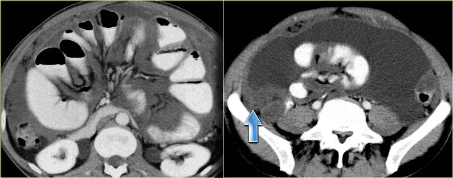
Paracolic Gutter Ct Anatomy
Paracolic Gutters

Inframesocolic Space Radiology Reference Article Radiopaedia Org

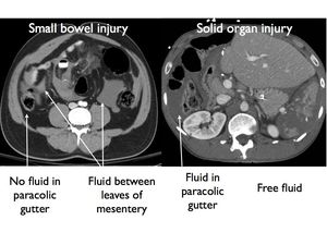
Ct Showing Free Fluid In The Right Paracolic Gutter No Free Air And Download Scientific Diagram
Http Pdf Posterng Netkey At Download Index Php Module Get Pdf By Id Poster Id 114921

Peritoneal Cavity Anatomy In Ct Peritoneography A Comprehensive Description Semantic Scholar
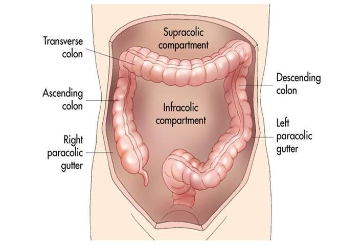
Paracolic Gutter

As the fluid continues superiorly into the inferior extension of the splenorenal recess and perhaps to some degree medially within the unusual variant of a retropancreatic recess.
Paracolic gutter ct anatomy.
Imaging Anatomy Of Peritoneum
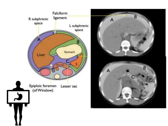
Peritoneum Intraperitoneal Spaces
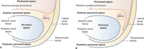
Abdomen And Pelvis Radiology Key
The Radiology Assistant Peritoneal Pathology
Http Pdf Posterng Netkey At Download Index Php Module Get Pdf By Id Poster Id 117837

Gastrointestinal Radiology
Left Paracolic Gutter
Http Pdf Posterng Netkey At Download Index Php Module Get Pdf By Id Poster Id 127176
Epos

Ct Showing Free Fluid In The Right Paracolic Gutter No Free Air And Download Scientific Diagram

Fitz Hugh Curtis Syndrome Radiology Case Radiopaedia Org
Http Pdf Posterng Netkey At Download Index Php Module Get Pdf By Id Poster Id 101511

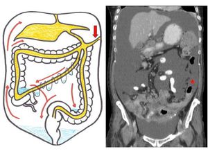
The Spread And Localization Of Intraperitoneal Abscesses Dynamic Radiology

Free Intraperitoneal Fluid Summary Radiology Reference Article Radiopaedia Org
Epos
Pericaecal Internal Hernia Radiology Case Radiopaedia Org

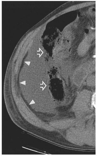
Pancreatic Injury Post Laparoscopic Subtotal Colectomy There Is Pancreatic Fluid Tracking From The Pancreatic Tail Arrows To Th Colectomy Abdomen Pancreatic

Peritoneal Carcinomatosis From Adenocarcinoma Of The Colon Ct Shows A Small Amount Of Ascites Nodular Soft Tissue Thick Disease Secondary Historical Figures
Https Encrypted Tbn0 Gstatic Com Images Q Tbn 3aand9gcqviiuoor7cqvjuy2tb0atddfdnjxh67jahtkkexb8 Usqp Cau

Ct Mri Upper Abdomen Normal Anatomy Dr Ahmed Eisawy Youtube

The Peritoneal Cavity Greater Sac Lesser Sac Teachmeanatomy

Jaypeedigital Ebook Reader
Abdomen Nontraumatic Emergencies Radiology Key
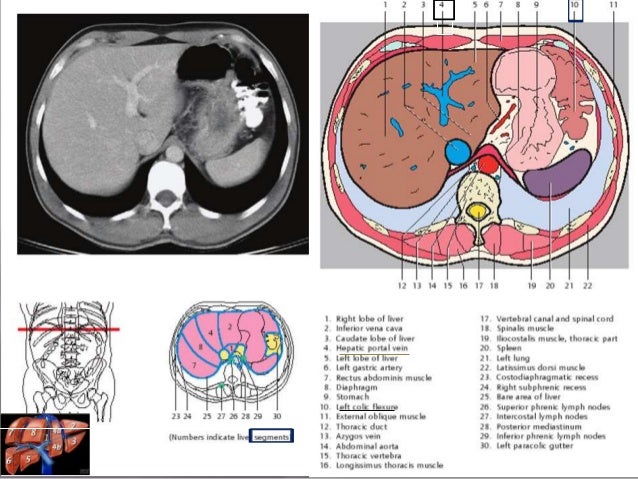
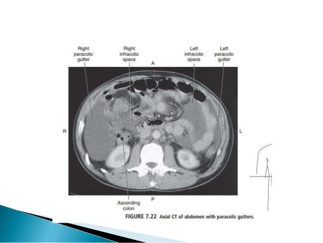
Sectional Anatomy Of Abdomen
Source : pinterest.com
